Moles In LOS GATOS, CA

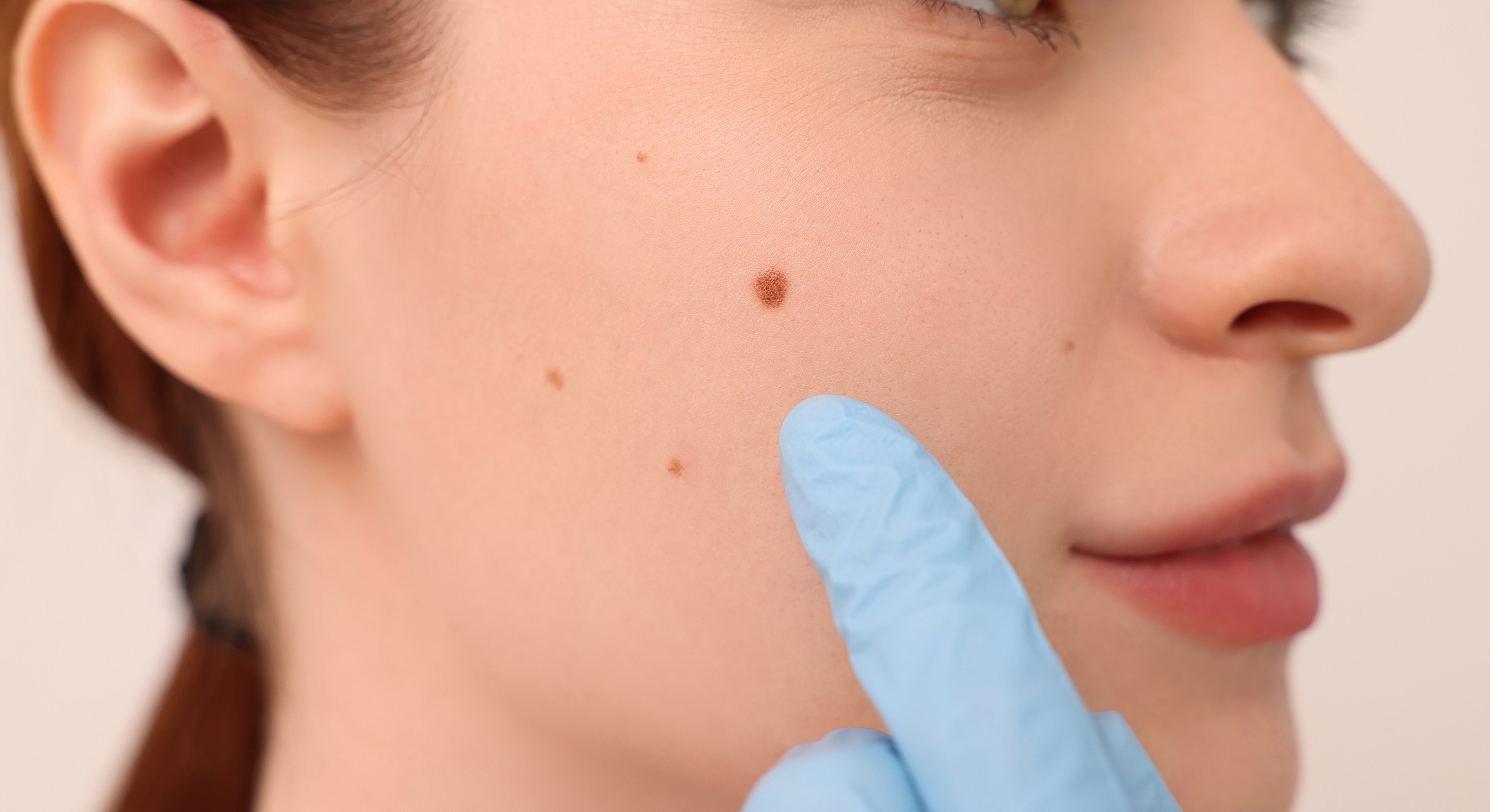
Moles, medically known as nevi, are common skin growths varying in size, shape, and color. These benign lesions develop from melanocytes, pigment-producing cells. Moles can appear anywhere, individually or in clusters, ranging from flat and light-colored to raised and darker. While most are harmless, understanding classifications like congenital and acquired nevi is crucial for monitoring changes.
The impact of moles is primarily aesthetic, though some may experience discomfort if a mole is in an area prone to friction. Beyond cosmetic concerns, the most significant impact relates to potential malignant transformation, particularly into melanoma. Regular self-examinations and professional dermatological screenings are essential for early detection and timely intervention.
Moles primarily result from clustered melanocytes, pigment-producing cells, forming a nevus. Genetic predisposition plays a significant role; individuals with a family history of moles or melanoma are more likely to develop them. Mole number and type are often attributed to inherited traits.
UV radiation from sunlight and tanning beds significantly contributes to new mole development and influences existing ones. UV exposure stimulates melanocyte proliferation and pigment production, increasing mole count and atypical change risk. Excessive sun exposure is a key environmental factor. Hormonal fluctuations during puberty or pregnancy can also alter existing moles or cause new ones.
Proactive mole prevention focuses on minimizing risk factors and diligent self-monitoring. Rigorous sun protection is critical, including consistent broad-spectrum sunscreen (SPF 30+), protective clothing, seeking shade, and avoiding tanning beds. Reducing UV exposure prevents new moles and protects existing ones from harmful changes.
Dr. Bitter's philosophy emphasizes proactive skin health and early intervention through regular skin examinations. Patients should perform monthly self-checks for new moles or changes in size, shape, color, or texture. Suspicious findings require prompt dermatological evaluation. Early detection of atypical moles or melanoma significantly improves treatment outcomes.
Dr. Patrick Bitter Jr., MD, is a world-renowned pioneer in aesthetic dermatology. He invented the FotoFacial™ in 1998, a groundbreaking procedure for non-invasive skin rejuvenation. His landmark 2000 study legitimized Intense Pulsed Light (IPL) as a powerful tool for treating various skin conditions.
Dr. Bitter's commitment to skin health is evidenced by his co-authorship of the 2013 Stanford gene expression study, proving Broadband Light (BBL) therapy reverses skin aging at a molecular level. With over 20,000 daily IPL/BBL procedures worldwide, his legacy as a global educator and innovator is unparalleled. Honored as an Aesthetic Medicine Icon in 2025, Dr. Bitter\'s expertise in distinguishing benign from concerning moles is paramount, and BBL treatments effectively address pigmented lesions.
BBL® HEROic™ is advanced Broadband Light therapy effectively targeting pigmented lesions, including certain moles. It delivers rapid, high-energy light pulses absorbed by melanin, leading to gradual fading and removal. This non-invasive solution improves skin tone and clarity, particularly for benign pigmented moles and sun-induced lesions.
HALO® TRIBRID™ is a hybrid fractional laser improving overall skin texture and tone, beneficial for moles. While not directly removing moles, HALO addresses surrounding skin irregularities and pigmentation, creating a more uniform complexion. It enhances the overall aesthetic outcome where moles are present.
Dr. Bitter's LaserSense™ line features Hypochlorous Acid (HOCl) products, rooted in the 2019 paper co-authored by Dr. Bitter, which established HOCl as the gold standard for wound care and scar management. This innovative line leverages HOCl\'s natural antimicrobial and anti- inflammatory properties to support skin healing and health. For moles, especially after removal procedures or for general skin health, these products are highly beneficial.
LaserSense™ Dermal Spray is recommended for its soothing and purifying properties, maintaining skin hygiene and reducing irritation in areas with moles. LaserSense™ Post Procedure Gel offers intensive skin recovery support, ideal after dermatological interventions for moles, promoting optimal healing and minimizing downtime.
Alastin Skincare® is renowned for its proprietary TriHex Technology®, a blend of active peptides and botanicals supporting the skin's natural ability to produce new, healthy elastin and collagen. This technology clears damaged proteins and replenishes skin, leading to a more youthful complexion. Maintaining overall skin health is crucial for individuals with moles.
Alastin Regenerating Skin Nectar is highly beneficial for preparing skin for procedures and supporting recovery, relevant if moles are monitored or removed. Alastin HydraTint Pro Mineral SPF 36 provides essential broad-spectrum sun protection, critical for preventing new moles and protecting existing ones from sun damage. Alastin A-LUMINATE Brightening Serum addresses surrounding hyperpigmentation or uneven skin tone, contributing to a more uniform appearance.
[1] Patrick Bitter Jr., MD. About. https://patrickbitterjrmd.com/about/
[2] Chang, A. L. S., et al. (2013). Noninvasive rejuvenation and volume augmentation of the face with broad-spectrum light and fractional resurfacing. PubMed. https://pubmed.ncbi.nlm.nih.gov/22931923/
[3] Gold, M. H., et al. (2016). Acne Treatment with 3-Step Broadband Light Protocol. Journal of Drugs in Dermatology. https://jddonline.com/articles/acne-treatment-with-3-step-broadband- light-protocol-S1545961616P1382X
[4] Bitter, P. H., & Pozner, J. N. (2019). Hypochlorous Acid: A Review of its Use in Wound Care and Scar Management. PubMed. https://pubmed.ncbi.nlm.nih.gov/31904191/
[5] Aesthetic Guide. (2026). From Peels to Protocols: The Modern Makeup of Cosmetic Dermatology. https://www.theaestheticguide.com/aesthetic-dermatology/from-peels-to- protocols-the-modern-makeup-of-cosmetic-dermatology
[6] Sciton. BBL HEROic: A Revolutionary Breakthrough in Aesthetics. https://sciton.com/blog-a- revolutionary-breakthrough-in-aesthetics-meet-the-worlds-smartest-pulsed-light-device-bbl- heroic/
